Tuesday, 12 March 2019

Cerebral Venous System


Cerebral Sinuses and Veins

There are 11 venous sinuses in total, between periostial and meningeal layers of dura.

Veins of significance include Thalamostriate, Superior Anastomotic Vein (of Trolard), Internal Cerebral Vein, Great Cerebral Vein (of Galen), Basal Vein (of Rosenthal), Inferior Anastomotic Vein (of Labbe), transverse vein, Middle Cerebral Veins (of Sylvius)- Deep and Superficial, Anterior cerebral vein, 
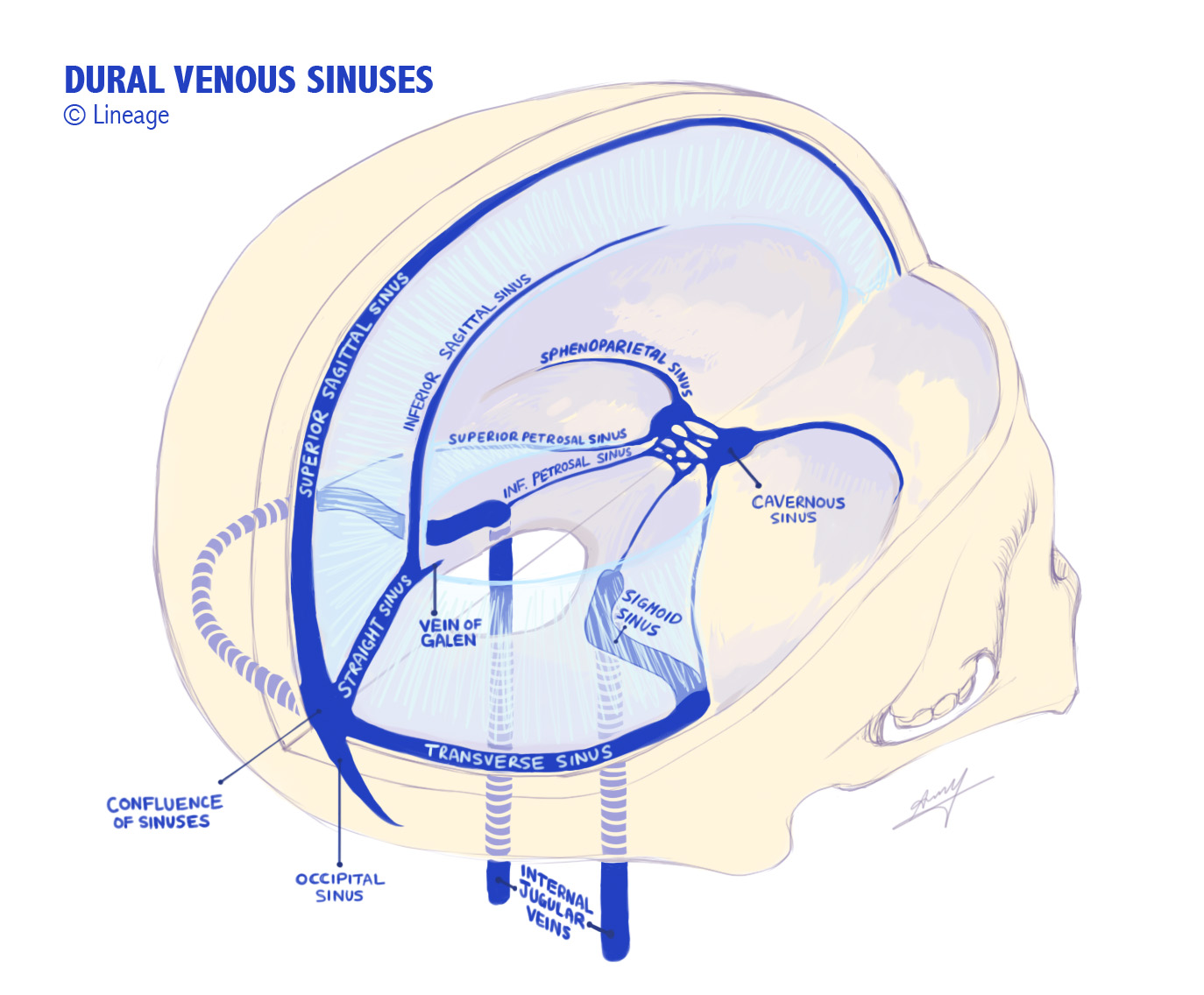
Image result for cerebral venous sinuses
Right transverse sinus is usually dominant and typically receives superficial blood- via continuation of SSS; left TS typically drains deep brain 


Venous Sinuses

NB inferior petrosal sinus sampling (to measure ATCH levels) is a clue that this drains pituitary.

Veins are located in subarachnoid space and divided into superficial and deep systems. 

Superficial Veins
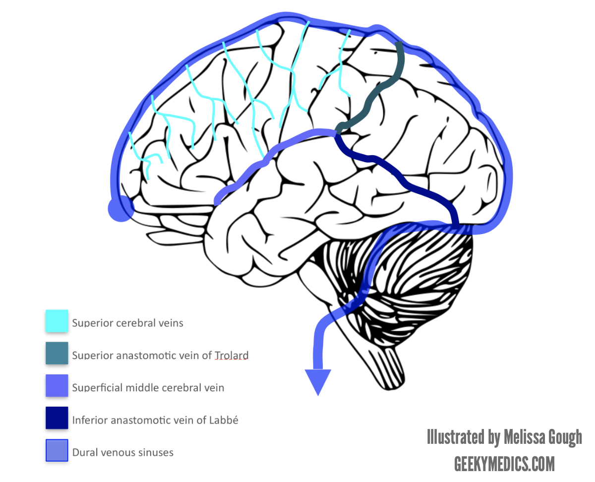
Image result for cerebral veins
Trolard, Labbe and Sylvius (superficial) all meet up.


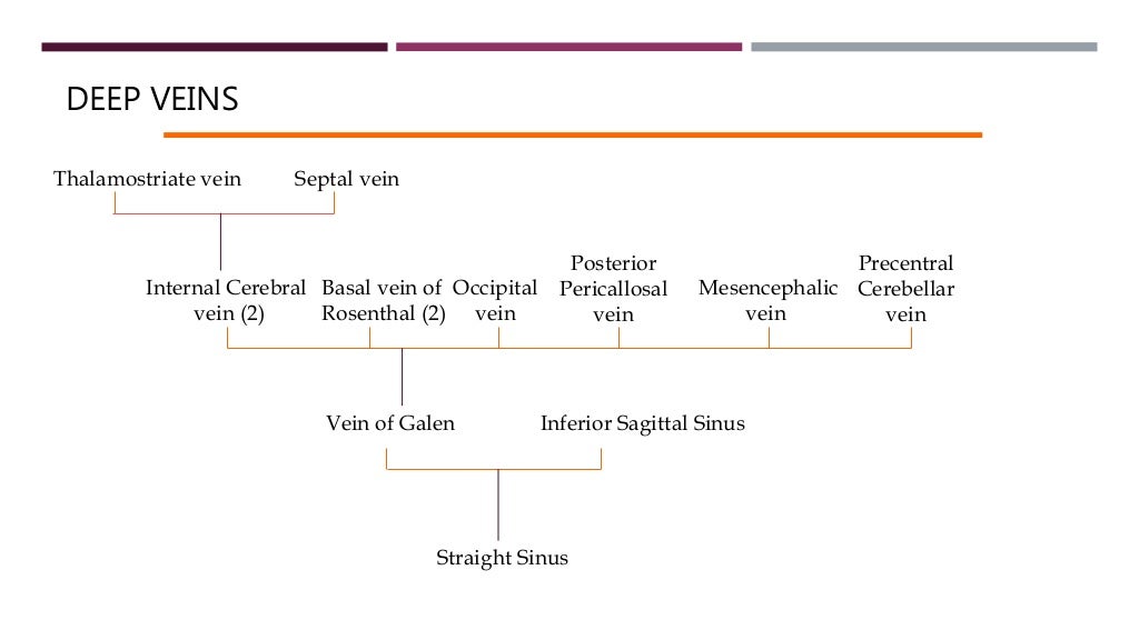
DEEP VEINSVEINS
Thalamostriate vein Septal vein
Internal Cerebral
vein (2)
Basal vein of
Rosenthal (2)
Occipital
vein
Post...


Visual Pathways

Visual Pathways 


Blue arrows indicate Meyer's Loop.


Optic nerve is developed from the optic vesicle. 
Originates from retinal ganglion cell axons, enters the cranial cavity via optical canal, passing through tendinous ring annulus of Zinn. 


Related image


"2, 3 n 6" pass within the annulus, n being nasociliary nerve (i.e. V1 branch).

Image result for optic radiation

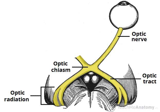
Meyer's loop is anterior, inferior and temporal; central bundle abuts lateral ventricle; the dorsal bundle is superior and parietal (Baum's loop). Meyer's loop produces pie in the sky deficit.

NB Nasal fibres cross at the chiasm, temporal don't (you cross your eyes at your nose).


This demonstrates the geniculate pathway conscious visual processing (incorporating the LGN of the thalamus)
There are also extrageniculate pathways, which project to: pretectal nucleus (midbrain- pupillary reflexes), superior colliculus (midbrain- coordinated reflexive head and eye movements), suprachiasmatic nucleus (hypothalamus- circadian cycles).

Image result for primary visual cortex


Sunday, 24 February 2019

Cranial Projection and Association Tracts and Limbic System


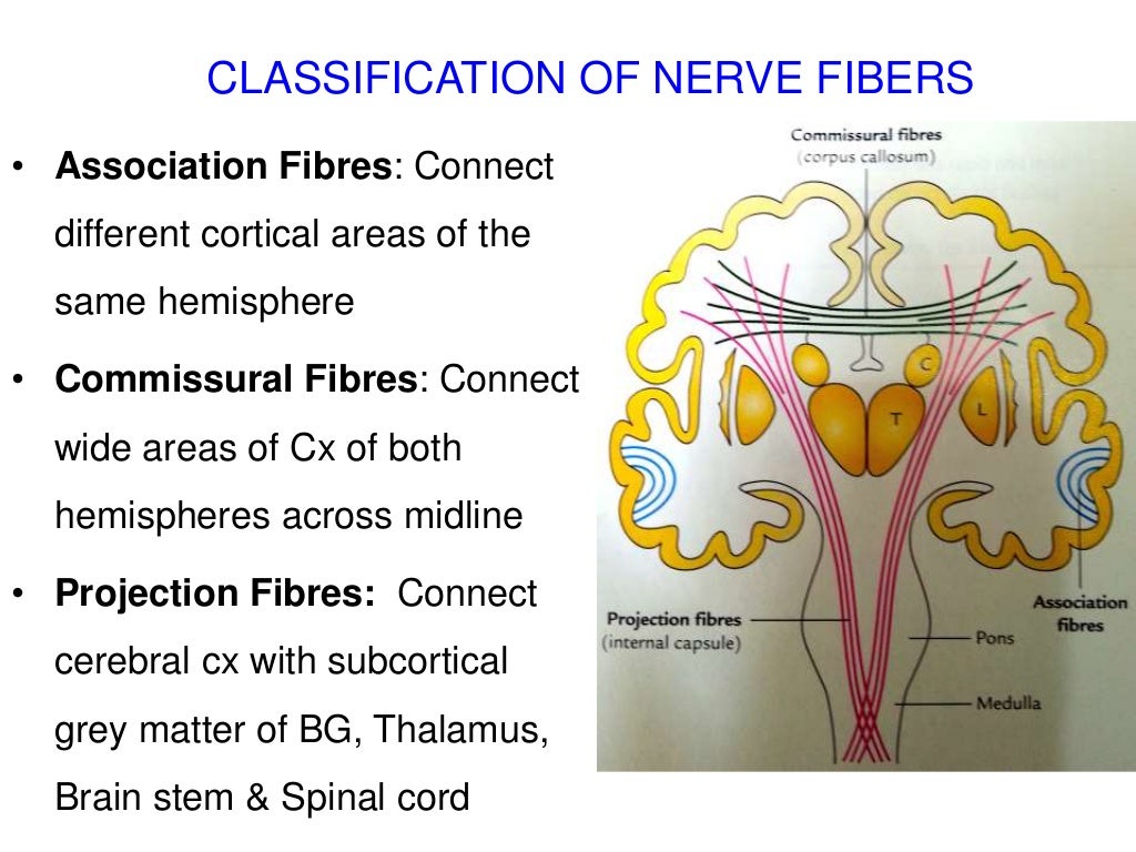
INTERNAL CAPSULE
• A compact band of
neocortical projection fibres
• Main highway for input &
output fibres of cerebral cx...


Image result for white matter association fibres


Bundles implies long association fibres

No photo description available.

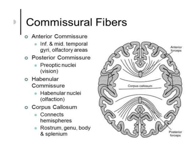
Image result for commissural fibresImage result for anterior commissure


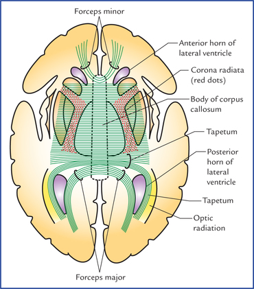
Corpus callosum fibres

Image result for tapetum corpus callosum


Image result for lentiform nucleus


Image result for lenticular fasciculus
VA- ventral anterior
VL - ventral lateral

Image result for lenticular fasciculus


NB Lentiform nucleus = GP + putamen

Tegmentum (Lat- Covering). Forms floor of midbrain cf.
Tectum (Lat- Roof). Forms roof of midbrain.
Stria (Lat- Stripe)
    Fasciculus (Lat- Bundle)
    Tapetum (Lat- Tapestry/rug)
    Habenula (Lat- reins). Denotes stalk of pineal gland and neighbouring neurons.


Image result for tegmentum


Resembles a tapestry; the carpet of the brain. Primarily formed by decussating fibres of the splenium.

Septal nuclei are by septum pellucidum. Role in pleaseure/reward like Nucleus Accumbus.


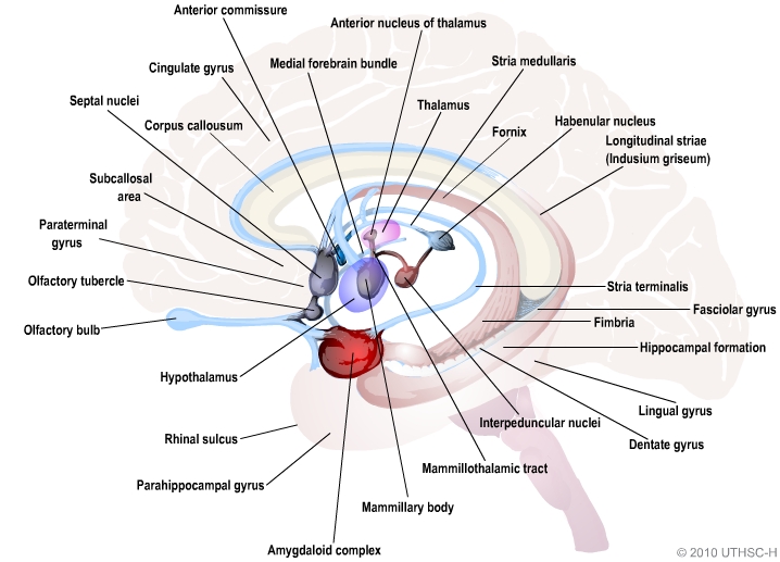
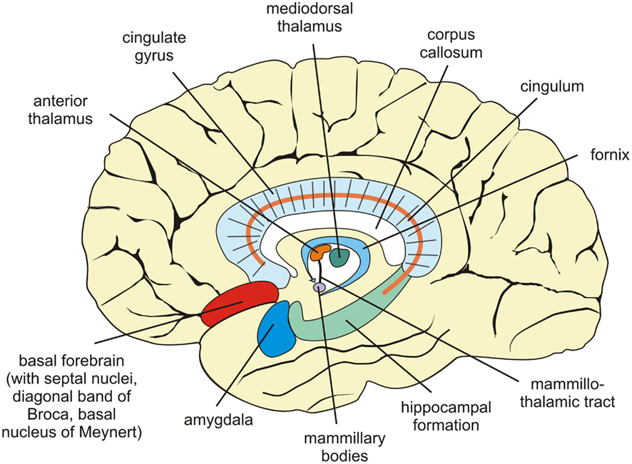
Limbic System- a ring of memory and emotion (like a wedding ring).


Image result for limbic system diagram sagittal


Simplified:

Image result for limbic system diagram sagittal

Image result for papez circuit

Deontes a conerstone of memory processing/consolidation


Indusium (tunic/shirt) griseum is membranous layer of grey matter which covers the corpus callosum.


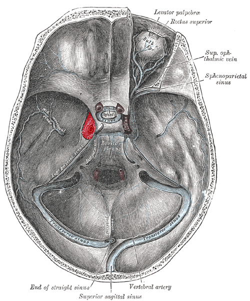
Cavernous Sinus

Cavernous Sinus Anatomy


All are in lateral wall except V1
From superior to inferior III, IV, V1, V2.


Image result for Cavernous sinus o tom cat


Coronal scan through the cavernos sinus. MR contrast-enhanced FIESTA sequence. a, oculomotor nerve; b, trochlear nerve; c, abducens nerve; d, ophthalmic nerve; e, maxillary nerve; f, internal cerebral artery; cavernous segment; g, right optic nerve; h, pituitary gland

Sunday, 27 January 2019

Middle Cerebral Artery


Middle Cerebral Artery:

Territory:

Image result for anterior cerebral artery territory

Supplies most of lateral surface of hemisphere, part of internal capsule (posterior part of anterior limb and anterior part of posterior limb) and basal ganglia.

Origin:


Segments:

-Frontal 2D view following
right vertebral artery
injection , there is a
normal appearance of the
cervical vertebral arter...

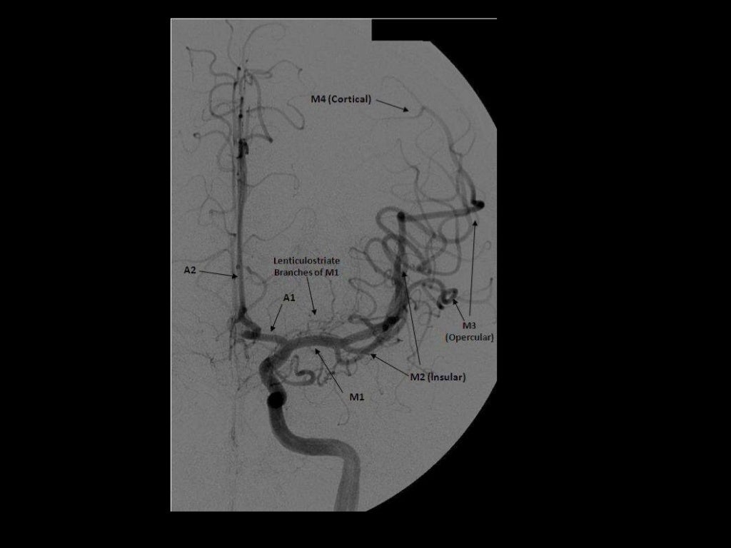
-2D frontal view following left ICA injections ,
these images show an aneurysm in the region
of the left MCA bifurcation/t...

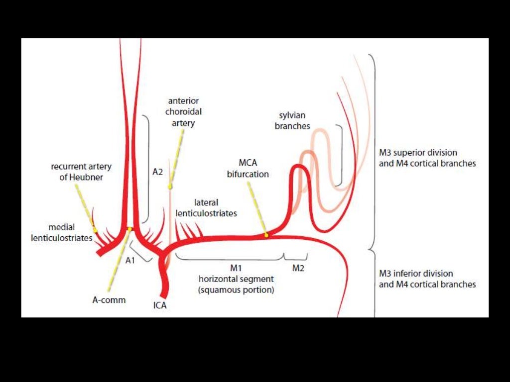
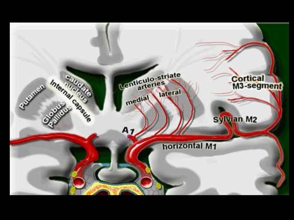
M1- ICA to Sylvian fissure (horizontal or Sphenoidal)
M2- Sylvian (within fissure; also Insular) post bifurcation into superior & inferior divisions. 
M3- Opercular: Top of fissure to cerebral cortex
M4- Cortical (cortical surface branches)


B-3D posterior view
following right
vertebral artery
injection , there is a
normal appearance of
the cervical vertebral
art...

Branches:
NB superficial and deep

M1
Lateral lenticulostriate A. (NB ACA is the more medial vessel)

M2 (NB superior and inferior divisions) 
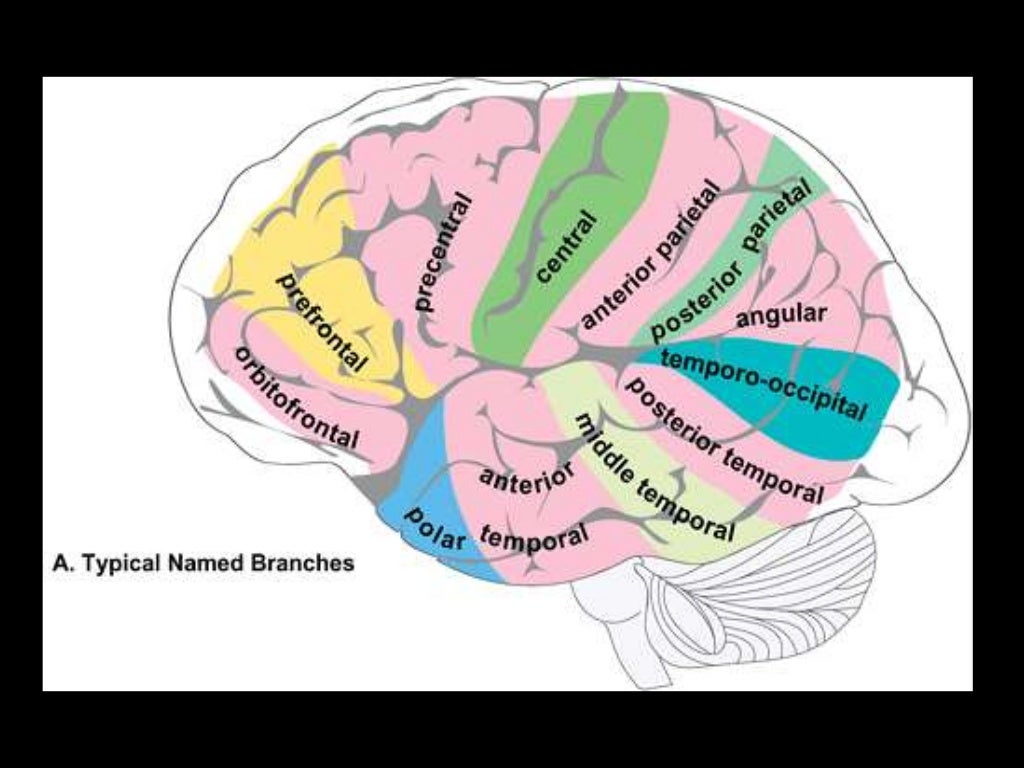
Branches supply temporal lobe and insular cortex, parietal lobe, inferolateral frontal lobe via:

Superior:
Lateral frontobasal A.
Prefrontal sulcas A.
Pre-Rolandic (Precentral) and Rolandic (central) sulcal A.

Inferior:
3 temporal branches (anterior, middle and posterior)
Branch to angular gyrus
2 parietal branches (anterior, posterior)
-3D frontal ) view following left
vertebral artery injection , this view
show intracranial vertebral basilar
circulation i...

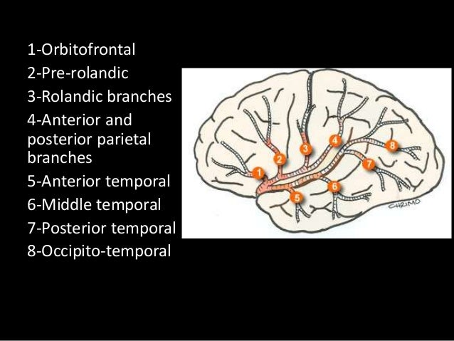
2-7 appear to be exclusively MCA branches. 1 shared with ACA. 8 PCA also.

-3D frontal view following right
vertebral artery injection , this
view shows normal intracranial
vertebral basilar circul...


M3 and M3
The above branches will have M3 and M4 portions


Anterior Cerebral Artery

Anterior Cerebral Artery

Territory:

Image result for anterior cerebral artery territory

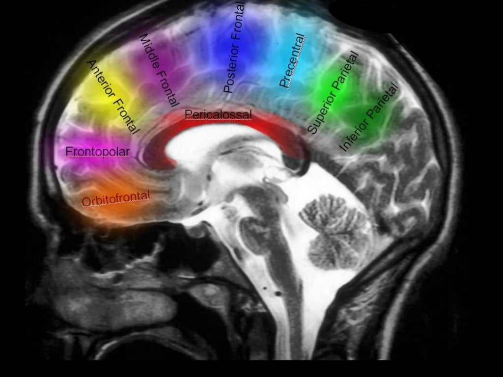
Supplies most of medial surface of cerebral cortex (anterior 3/4), frontal pole and anterior portions of corpus callosum.
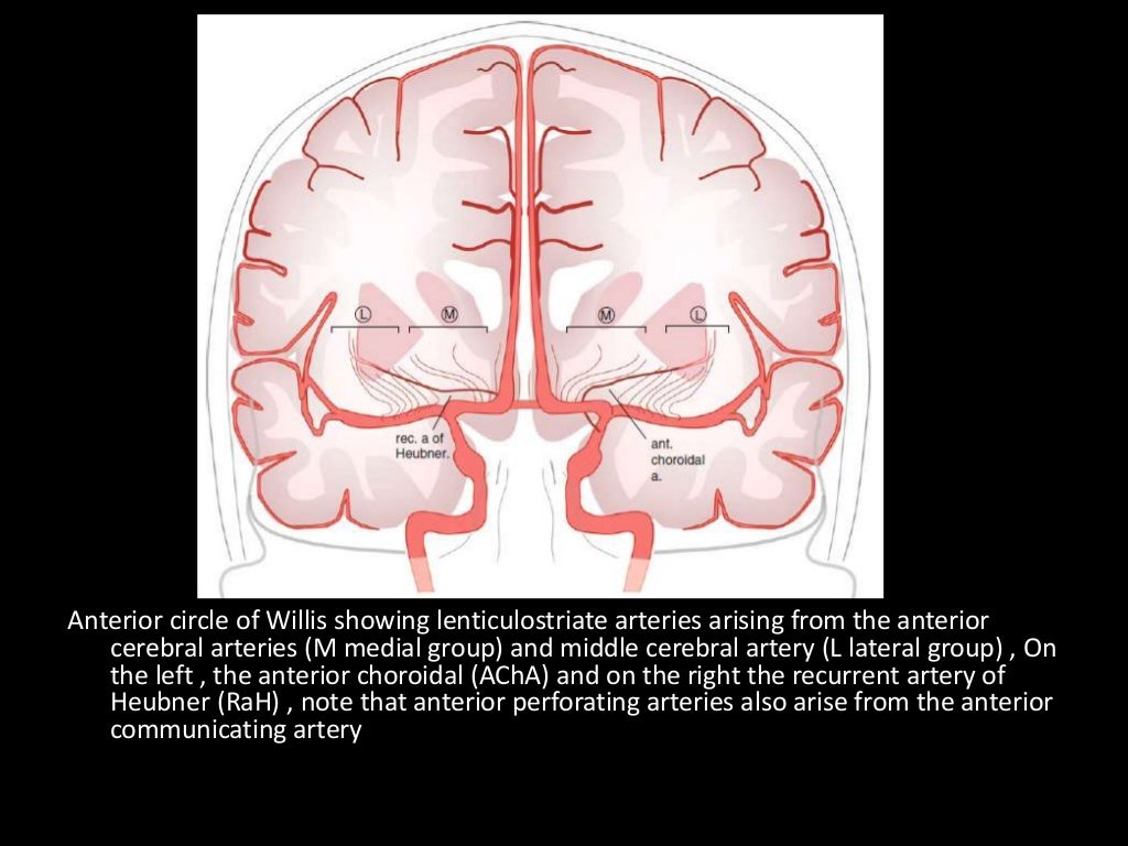
Perforating branches supply anterior limb of the internal capsule, inferior portions of caudate head and anterior globus pallidus.

Infarction will potentially cause (mainly) lower limb weakness, incontinence, abulia/motor aphasia, frontal lobe symptoms.


Origin
Related image

Segments

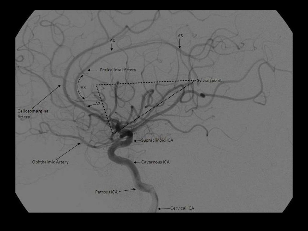
A1 is the segment originating from the internal carotid artery and extending to the anterior communicating artery. A2 extends from the anterior communicating artery to a region between the rostrum and genu. The A3 segment courses around the genu to the rostral part of the body. A4 and A5 segments are the continuation of the pericallosal artery. 

A1: ICA bifurcation to Acomm (horizontal)
A2: Acomm to origin of CMA around rostrum/genu junction (vertical)
A3: Genu to body of CC (proximal pericallosal)
A4/A5: Continuation of distal pericallosal (directly posterior)

(1) MCA (M4) , (2) MCA (M3) , (3) MCA (M2) , (4) MCA (M1) , (5) LT ICA , (6) LT
MCA , (7) Basilar artery , (8) PCA

NB angiographic sylvian point = apex of fissure sf. skull sylvian point a surface marking for fissure 3cm behind superior portion of zygomatic process


anteriorcerebralartery.jpg

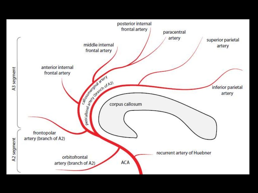
Branches

A1
Medial lenticulostriate A.
AComm A.

A2
Recurrent artery of Hubner (= medial striate A.). Supplies head of caudate and anterior limb of IC. (Can be A1 branch)
Orbitofrontal A.
Frontopolar A.

A3
Callosomarginal A. (in cingulate sulcus)
Pericallosal A.
Anterior (internal) frontal A.
Middle (internal) frontal A.
Posterior (internal) frontal A.
Paracentral A.


NB Striatum =  Caudate + IC + lentiform nucleus (which = putamen + GP).

a) Deep (Perforating) Branches :
-Arise from the superior surface of the M1
segment
-They are grouped as the medial & late...

-2D frontal view following left ICA injections ,
these images show an aneurysm in the region
of the left MCA bifurcation/t...


*Arteries to the Parietal & Occipital lobes:
-These run posterior to the sylvian fissure , from
superior to inferior :
1-A...

1-Orbitofrontal , 2-Prefrontal , 3-Precentral , 4-Central , 5-Anterior parietal , 6-Post
parietal , 7-Angular , 8-Occipito...

Sunday, 2 December 2018