Thursday 14 March 2019

Thalamic Nuclei

Thalamic Nuclei

Image result for thalamic nuclei

If you get orientated, they are largely as they say on the tin
Pulvinar is posterior (means cushion- you sit on it with your posterior)
The ventral 'belly' side faces outwards *laterally).

Image result for thalamic nuclei


Image result for thalamic nuclei

Wednesday 13 March 2019

Parasympathetics

Parasympathetics


FeatureSympathetic NSParasympathetic NS
Summary of responsesFight or flightRest and digest
Spinal cord distributionThoracolumbarCraniosacral
Preganglionic neuroneShortLong
Preganglionic neurotransmitterAcetylcholine (Ach, cholinergic)Acetylcholine (Ach, cholinergic)
Postganglionic neuroneLongShort
Postganglionic neurotransmitterNoradrenaline (NA, adrenergic) in most cases*Acetylcholine (Ach, cholinergic)

*Sweat being an exception 

Efferent i.e. motor
Afferent i.e. towards the CNS so sensory 

Parasympathetic motor efferents—This system is divided into cranial and sacral components, and parasympathetic efferents only synapse with postganglionic cells close to or within target viscera (allowing local discrete responses)

4 main parasympathetic nuclei (III, VII, IX, X) with synapses in 4 ganglia (NB ganglia are outside CNS)


NB Most ganglia are associated with some sensory and sympathetic nerves – these do not synapse in the ganglia, they merely travel through it. 

1. Cilliary (parasympathetic eye) 

Supplied by fibres from Edinger-Westphal nucleus (a/w CNIII) 
Function: pupil constriction and accommodation 

Image result for ciliary ganglion

2. Pterygopalatine Ganglion (aka sphenopalatine) in pterygopalatine fossa (cries/lubricates nasopharynx) 

Supplied by superior salivatory nuclues (a/w CN VII)  via GSPN and Vidian nerve. 
Function: secretomotor innervation to lacrimal gland, mucous glands of nasal cavity, nasopharynx and palate. 

Image result for pterygopalatine ganglion

3. Submandibular ganglion (salivates) 

Suspended from lingual nerve 
Supplied by superior salivatory nucleus (a/w CN VII) 
Function: Secretomotor innervation to the submandibular and sublingual salivary glands 

Image result for submandibular ganglion

4. OTic Ganglion (parOTid - salivates) 
Inferior to foramen ovale 
Supplied by inferior salivatory nuclues (a/w CN IX)
Innervates parotid 

Image result for otic ganglion


NB Vagus (X) does not innervate head and neck structures - only thorax and abdomen. 

NucleusPre-ganglionicGanglionPost-ganglionicTarget organs
Edinger-Westphal (Oculomotor nerve)Travels with the motor root of the oculomotor nerveCiliary ganglionTravels via the short ciliary nervesSphincter pupilliae
Ciliary muscles
Superior salivatory nucleus (Facial nerve)

NB VII above IX hence sup and inf (both salivate) 
Travels with the greater petrosal nerve and the nerve of the pterygoid canalPterygopalatine ganglionHitchhikes on branches of the maxillary nerveLacrimal gland
Nasopharynx
Palate
Nasal cavity
Travels within the chorda tympani, a branch of the facial nerveSubmandibular ganglionFibres travel directly to target organsSublingual and submandibular glands
Inferior salivatory nucleus (Glossopharyngeal nerve)Travels within the lesser petrosal nerveOtic ganglionHitchhikes on the auriculotemporal nerveParotid gland
Dorsal vagal motor nucleus (vagus nerve)Travels within the vagus nerveMany – located within the target organsn/aSmooth muscle of the trachea, bronchi and gastro-intestinal tract

Tuesday 12 March 2019

Cerebral Venous System


Cerebral Sinuses and Veins

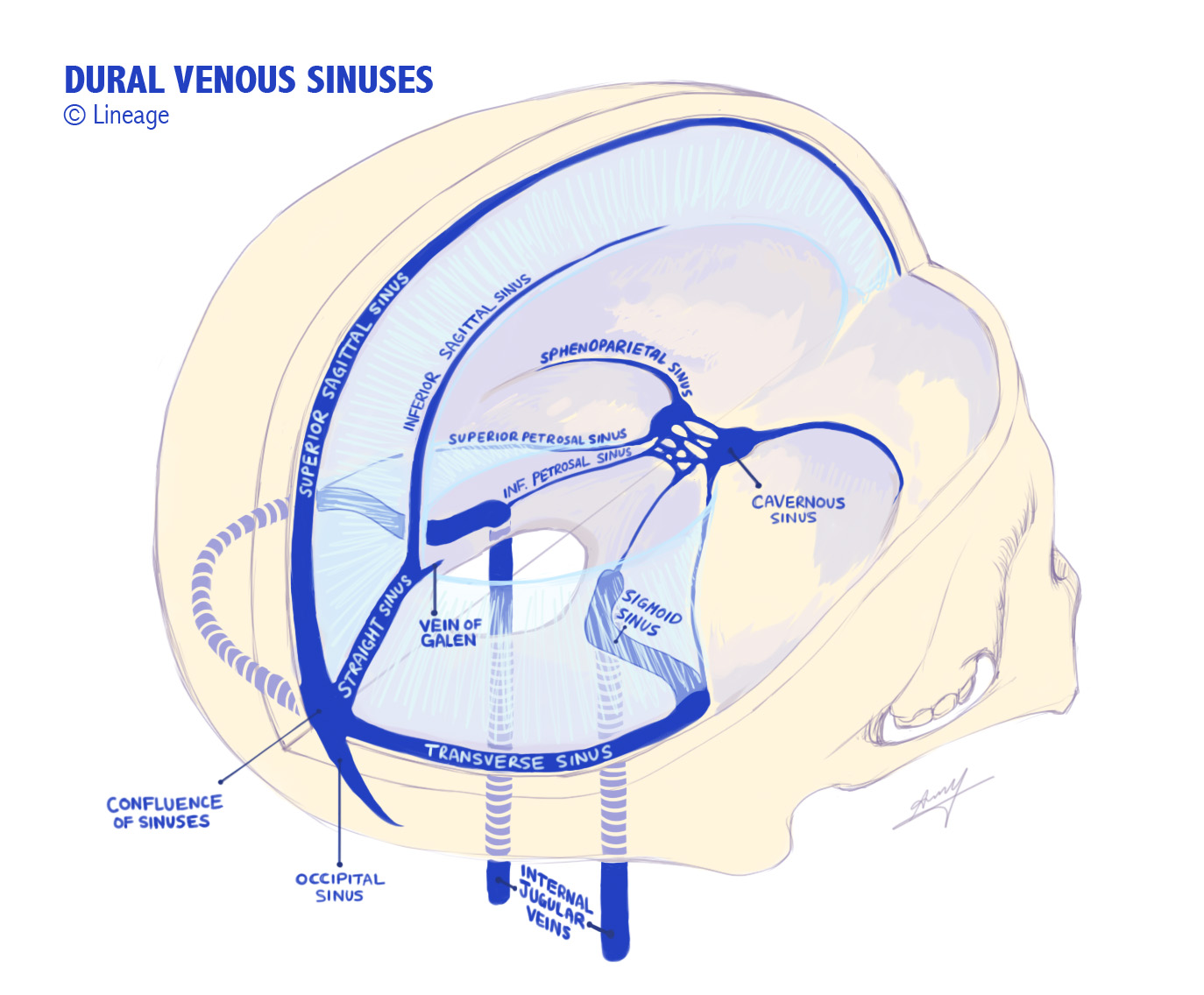
There are 11 venous sinuses in total, between periostial and meningeal layers of dura.

Veins of significance include Thalamostriate, Superior Anastomotic Vein (of Trolard), Internal Cerebral Vein, Great Cerebral Vein (of Galen), Basal Vein (of Rosenthal), Inferior Anastomotic Vein (of Labbe), transverse vein, Middle Cerebral Veins (of Sylvius)- Deep and Superficial, Anterior cerebral vein, 

Image result for cerebral venous sinuses
Right transverse sinus is usually dominant and typically receives superficial blood- via continuation of SSS; left TS typically drains deep brain 


Venous Sinuses

NB inferior petrosal sinus sampling (to measure ATCH levels) is a clue that this drains pituitary.

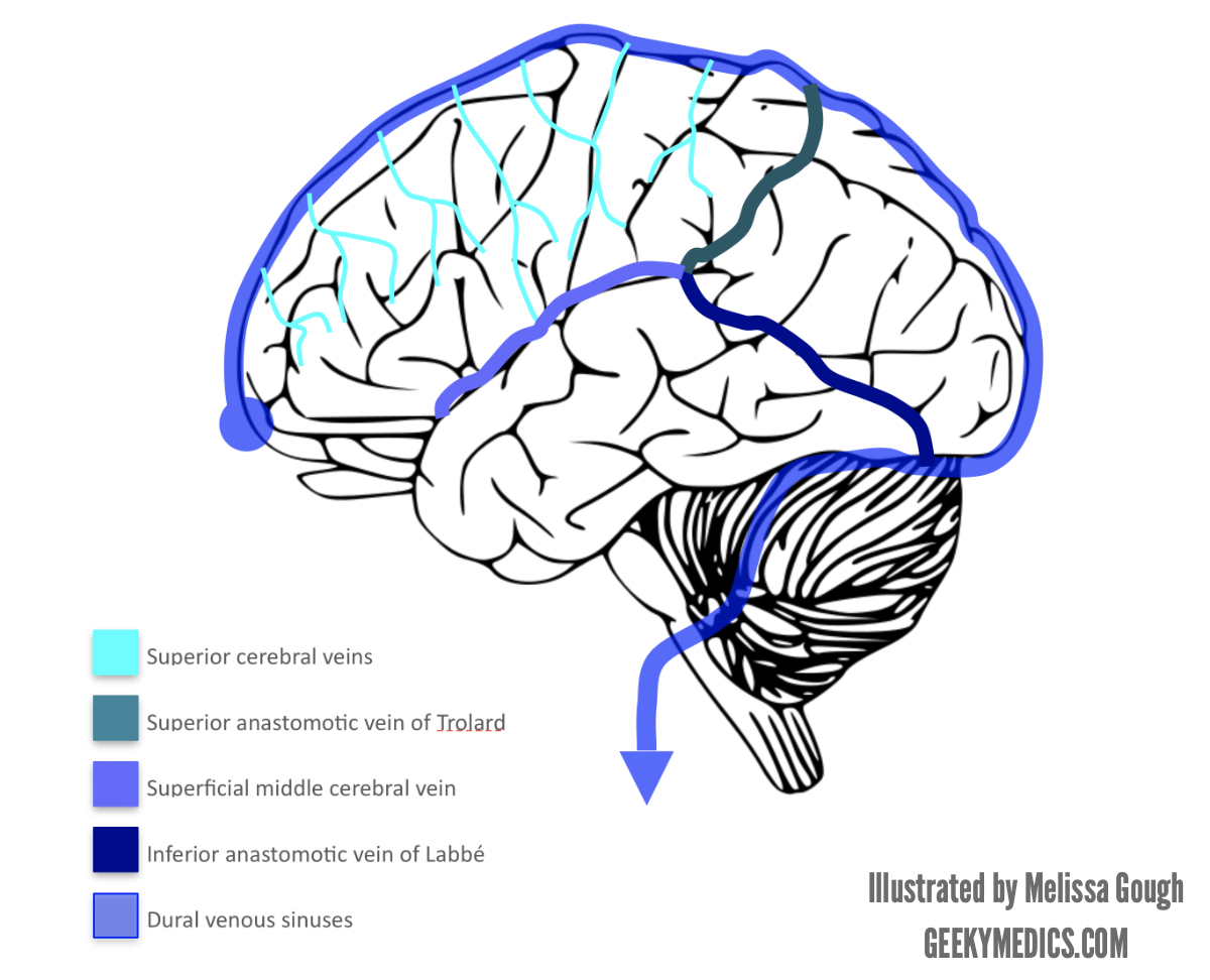
Veins are located in subarachnoid space and divided into superficial and deep systems. 

Superficial Veins

Image result for cerebral veins
Trolard, Labbe and Sylvius (superficial) all meet up.


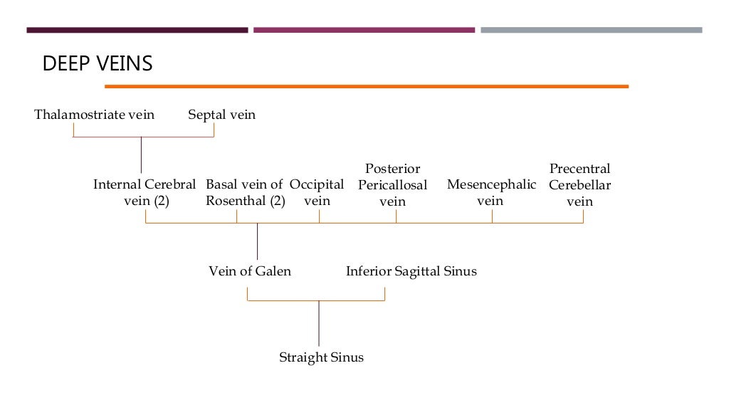
DEEP VEINSVEINS
Thalamostriate vein Septal vein
Internal Cerebral
vein (2)
Basal vein of
Rosenthal (2)
Occipital
vein
Post...


Visual Pathways

Visual Pathways 


Blue arrows indicate Meyer's Loop.


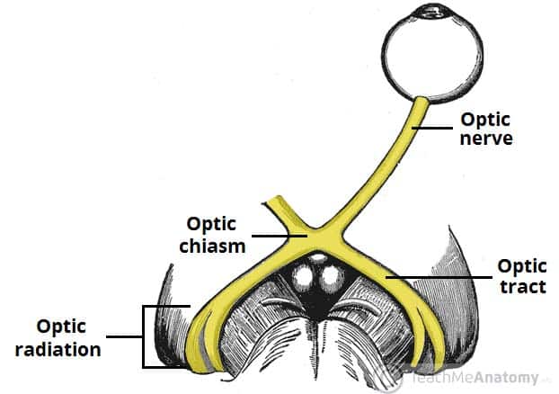
Optic nerve is developed from the optic vesicle. 
Originates from retinal ganglion cell axons, enters the cranial cavity via optical canal, passing through tendinous ring annulus of Zinn. 


Related image


"2, 3 n 6" pass within the annulus, n being nasociliary nerve (i.e. V1 branch).

Image result for optic radiation

Meyer's loop is anterior, inferior and temporal; central bundle abuts lateral ventricle; the dorsal bundle is superior and parietal (Baum's loop). Meyer's loop produces pie in the sky deficit.

NB Nasal fibres cross at the chiasm, temporal don't (you cross your eyes at your nose).


This demonstrates the geniculate pathway conscious visual processing (incorporating the LGN of the thalamus)
There are also extrageniculate pathways, which project to: pretectal nucleus (midbrain- pupillary reflexes), superior colliculus (midbrain- coordinated reflexive head and eye movements), suprachiasmatic nucleus (hypothalamus- circadian cycles).

Image result for primary visual cortex