Sunday 27 January 2019

Middle Cerebral Artery


Middle Cerebral Artery:

Territory:

Image result for anterior cerebral artery territory

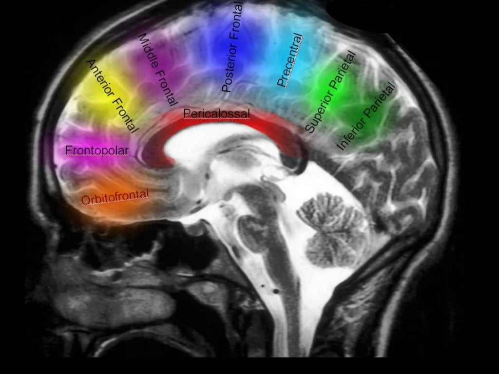
Supplies most of lateral surface of hemisphere, part of internal capsule (posterior part of anterior limb and anterior part of posterior limb) and basal ganglia.

Origin:


Segments:

-Frontal 2D view following
right vertebral artery
injection , there is a
normal appearance of the
cervical vertebral arter...

-2D frontal view following left ICA injections ,
these images show an aneurysm in the region
of the left MCA bifurcation/t...

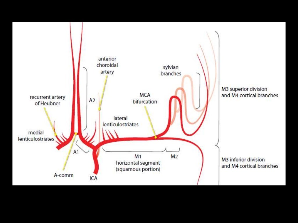
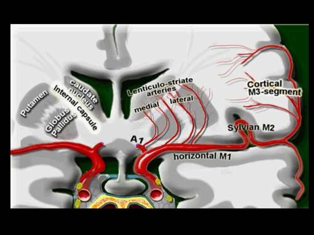
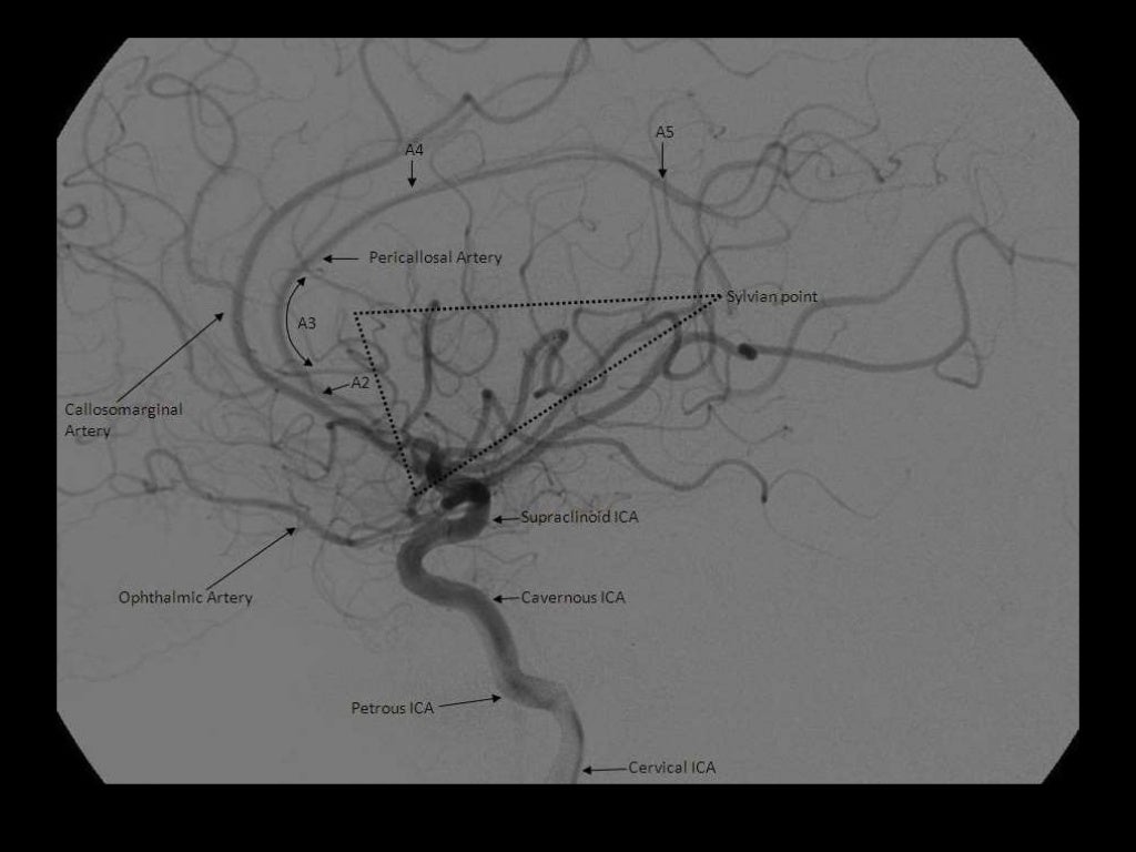
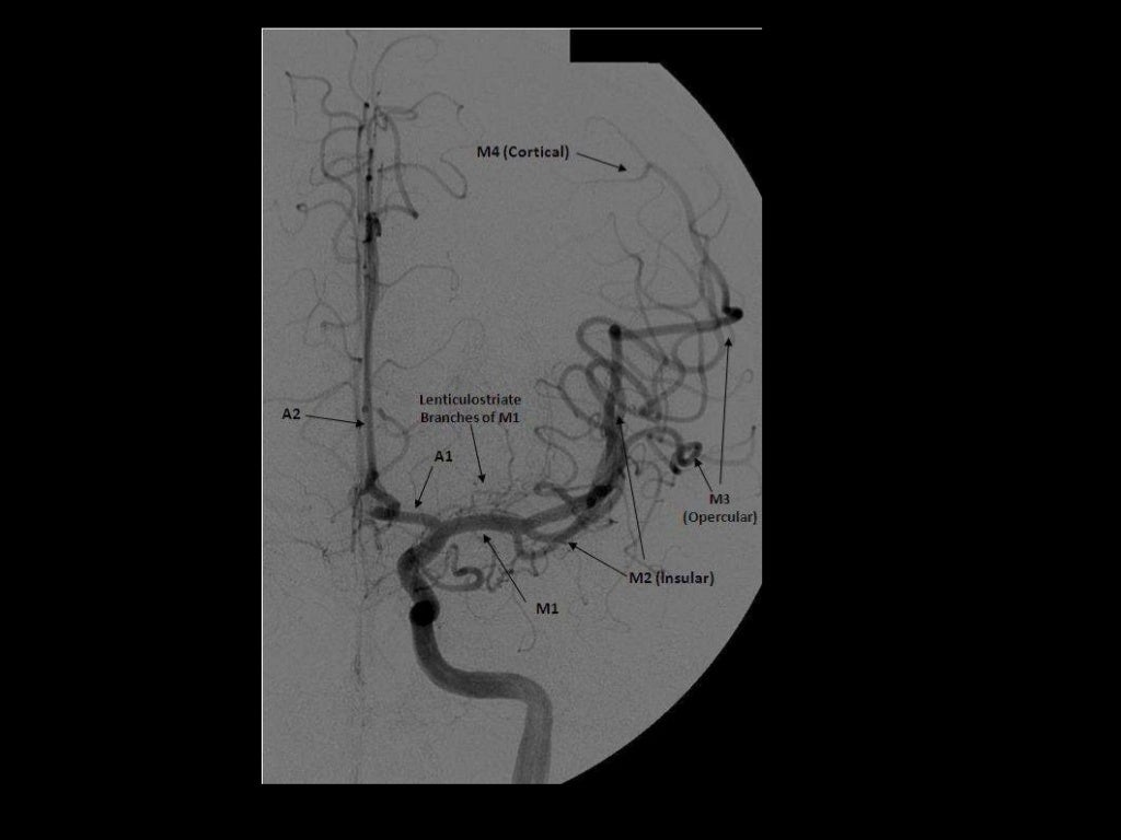
M1- ICA to Sylvian fissure (horizontal or Sphenoidal)
M2- Sylvian (within fissure; also Insular) post bifurcation into superior & inferior divisions. 
M3- Opercular: Top of fissure to cerebral cortex
M4- Cortical (cortical surface branches)


B-3D posterior view
following right
vertebral artery
injection , there is a
normal appearance of
the cervical vertebral
art...

Branches:
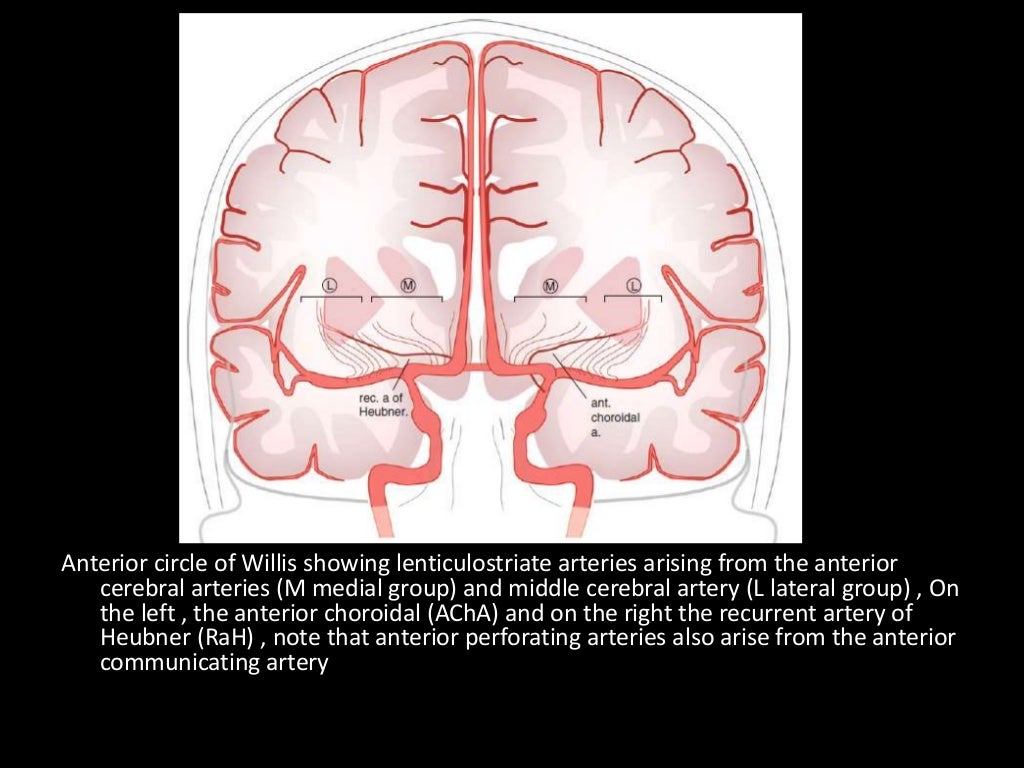
NB superficial and deep

M1
Lateral lenticulostriate A. (NB ACA is the more medial vessel)

M2 (NB superior and inferior divisions) 
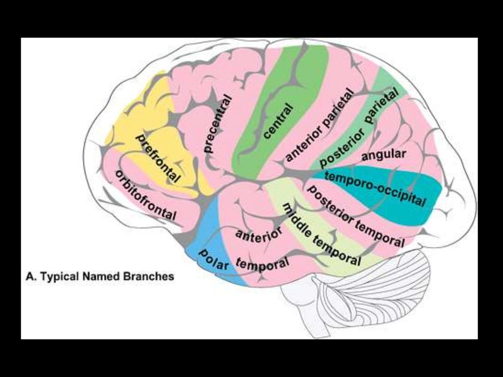
Branches supply temporal lobe and insular cortex, parietal lobe, inferolateral frontal lobe via:

Superior:
Lateral frontobasal A.
Prefrontal sulcas A.
Pre-Rolandic (Precentral) and Rolandic (central) sulcal A.

Inferior:
3 temporal branches (anterior, middle and posterior)
Branch to angular gyrus
2 parietal branches (anterior, posterior)
-3D frontal ) view following left
vertebral artery injection , this view
show intracranial vertebral basilar
circulation i...

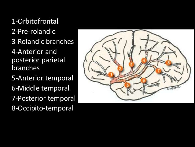
2-7 appear to be exclusively MCA branches. 1 shared with ACA. 8 PCA also.

-3D frontal view following right
vertebral artery injection , this
view shows normal intracranial
vertebral basilar circul...


M3 and M3
The above branches will have M3 and M4 portions


Anterior Cerebral Artery

Anterior Cerebral Artery

Territory:

Image result for anterior cerebral artery territory

Supplies most of medial surface of cerebral cortex (anterior 3/4), frontal pole and anterior portions of corpus callosum.
Perforating branches supply anterior limb of the internal capsule, inferior portions of caudate head and anterior globus pallidus.

Infarction will potentially cause (mainly) lower limb weakness, incontinence, abulia/motor aphasia, frontal lobe symptoms.


Origin
Related image

Segments

A1 is the segment originating from the internal carotid artery and extending to the anterior communicating artery. A2 extends from the anterior communicating artery to a region between the rostrum and genu. The A3 segment courses around the genu to the rostral part of the body. A4 and A5 segments are the continuation of the pericallosal artery. 

A1: ICA bifurcation to Acomm (horizontal)
A2: Acomm to origin of CMA around rostrum/genu junction (vertical)
A3: Genu to body of CC (proximal pericallosal)
A4/A5: Continuation of distal pericallosal (directly posterior)

(1) MCA (M4) , (2) MCA (M3) , (3) MCA (M2) , (4) MCA (M1) , (5) LT ICA , (6) LT
MCA , (7) Basilar artery , (8) PCA

NB angiographic sylvian point = apex of fissure sf. skull sylvian point a surface marking for fissure 3cm behind superior portion of zygomatic process


anteriorcerebralartery.jpg

Branches

A1
Medial lenticulostriate A.
AComm A.

A2
Recurrent artery of Hubner (= medial striate A.). Supplies head of caudate and anterior limb of IC. (Can be A1 branch)
Orbitofrontal A.
Frontopolar A.

A3
Callosomarginal A. (in cingulate sulcus)
Pericallosal A.
Anterior (internal) frontal A.
Middle (internal) frontal A.
Posterior (internal) frontal A.
Paracentral A.


NB Striatum =  Caudate + IC + lentiform nucleus (which = putamen + GP).

a) Deep (Perforating) Branches :
-Arise from the superior surface of the M1
segment
-They are grouped as the medial & late...

-2D frontal view following left ICA injections ,
these images show an aneurysm in the region
of the left MCA bifurcation/t...


*Arteries to the Parietal & Occipital lobes:
-These run posterior to the sylvian fissure , from
superior to inferior :
1-A...

1-Orbitofrontal , 2-Prefrontal , 3-Precentral , 4-Central , 5-Anterior parietal , 6-Post
parietal , 7-Angular , 8-Occipito...