
Blue arrows indicate Meyer's Loop.

Optic nerve is developed from the optic vesicle.
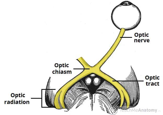
Originates from retinal ganglion cell axons, enters the cranial cavity via optical canal, passing through tendinous ring annulus of Zinn.

"2, 3 n 6" pass within the annulus, n being nasociliary nerve (i.e. V1 branch).

Meyer's loop is anterior, inferior and temporal; central bundle abuts lateral ventricle; the dorsal bundle is superior and parietal (Baum's loop). Meyer's loop produces pie in the sky deficit.
NB Nasal fibres cross at the chiasm, temporal don't (you cross your eyes at your nose).


This demonstrates the geniculate pathway conscious visual processing (incorporating the LGN of the thalamus)
There are also extrageniculate pathways, which project to: pretectal nucleus (midbrain- pupillary reflexes), superior colliculus (midbrain- coordinated reflexive head and eye movements), suprachiasmatic nucleus (hypothalamus- circadian cycles).

No comments:
Post a Comment