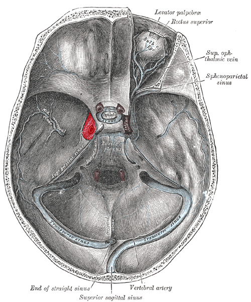


All are in lateral wall except V1
From superior to inferior III, IV, V1, V2.


Coronal scan through the cavernos sinus. MR contrast-enhanced FIESTA sequence. a, oculomotor nerve; b, trochlear nerve; c, abducens nerve; d, ophthalmic nerve; e, maxillary nerve; f, internal cerebral artery; cavernous segment; g, right optic nerve; h, pituitary gland
No comments:
Post a Comment