Sunday, 24 February 2019

Cavernous Sinus

Cavernous Sinus Anatomy


All are in lateral wall except V1
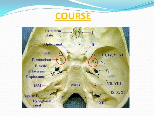
From superior to inferior III, IV, V1, V2.


Image result for Cavernous sinus o tom cat


Coronal scan through the cavernos sinus. MR contrast-enhanced FIESTA sequence. a, oculomotor nerve; b, trochlear nerve; c, abducens nerve; d, ophthalmic nerve; e, maxillary nerve; f, internal cerebral artery; cavernous segment; g, right optic nerve; h, pituitary gland

Sunday, 27 January 2019

Middle Cerebral Artery


Middle Cerebral Artery:

Territory:

Image result for anterior cerebral artery territory

Supplies most of lateral surface of hemisphere, part of internal capsule (posterior part of anterior limb and anterior part of posterior limb) and basal ganglia.

Origin:


Segments:

-Frontal 2D view following
right vertebral artery
injection , there is a
normal appearance of the
cervical vertebral arter...

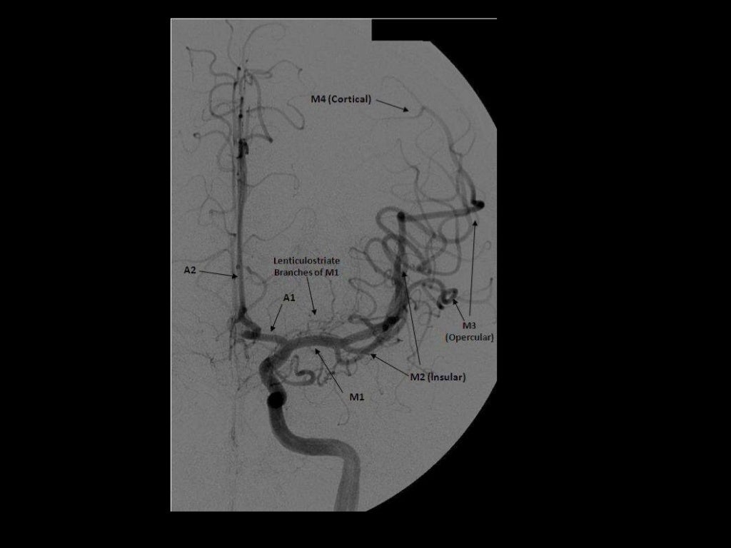
-2D frontal view following left ICA injections ,
these images show an aneurysm in the region
of the left MCA bifurcation/t...

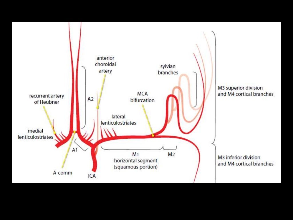
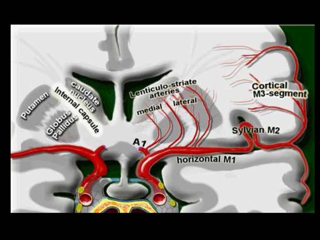
M1- ICA to Sylvian fissure (horizontal or Sphenoidal)
M2- Sylvian (within fissure; also Insular) post bifurcation into superior & inferior divisions. 
M3- Opercular: Top of fissure to cerebral cortex
M4- Cortical (cortical surface branches)


B-3D posterior view
following right
vertebral artery
injection , there is a
normal appearance of
the cervical vertebral
art...

Branches:
NB superficial and deep

M1
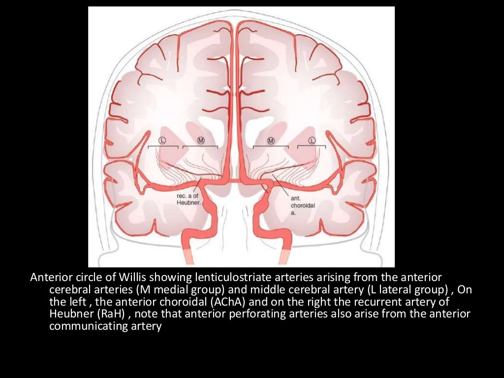
Lateral lenticulostriate A. (NB ACA is the more medial vessel)

M2 (NB superior and inferior divisions) 
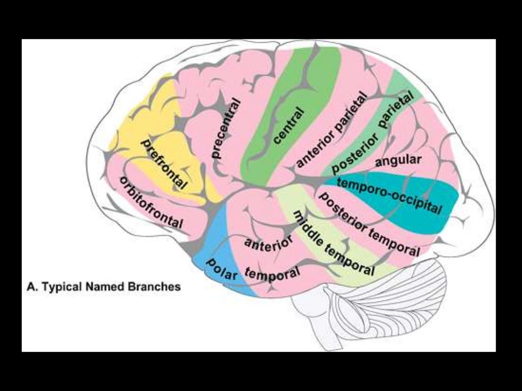
Branches supply temporal lobe and insular cortex, parietal lobe, inferolateral frontal lobe via:

Superior:
Lateral frontobasal A.
Prefrontal sulcas A.
Pre-Rolandic (Precentral) and Rolandic (central) sulcal A.

Inferior:
3 temporal branches (anterior, middle and posterior)
Branch to angular gyrus
2 parietal branches (anterior, posterior)
-3D frontal ) view following left
vertebral artery injection , this view
show intracranial vertebral basilar
circulation i...

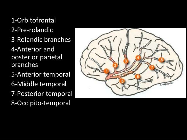
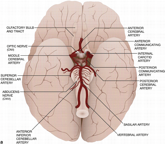
2-7 appear to be exclusively MCA branches. 1 shared with ACA. 8 PCA also.

-3D frontal view following right
vertebral artery injection , this
view shows normal intracranial
vertebral basilar circul...


M3 and M3
The above branches will have M3 and M4 portions


Anterior Cerebral Artery

Anterior Cerebral Artery

Territory:

Image result for anterior cerebral artery territory

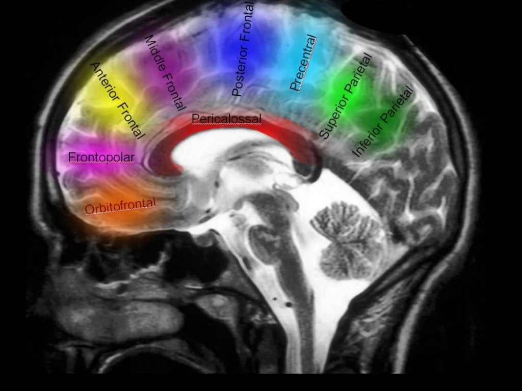
Supplies most of medial surface of cerebral cortex (anterior 3/4), frontal pole and anterior portions of corpus callosum.
Perforating branches supply anterior limb of the internal capsule, inferior portions of caudate head and anterior globus pallidus.

Infarction will potentially cause (mainly) lower limb weakness, incontinence, abulia/motor aphasia, frontal lobe symptoms.


Origin
Related image

Segments

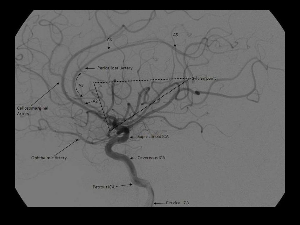
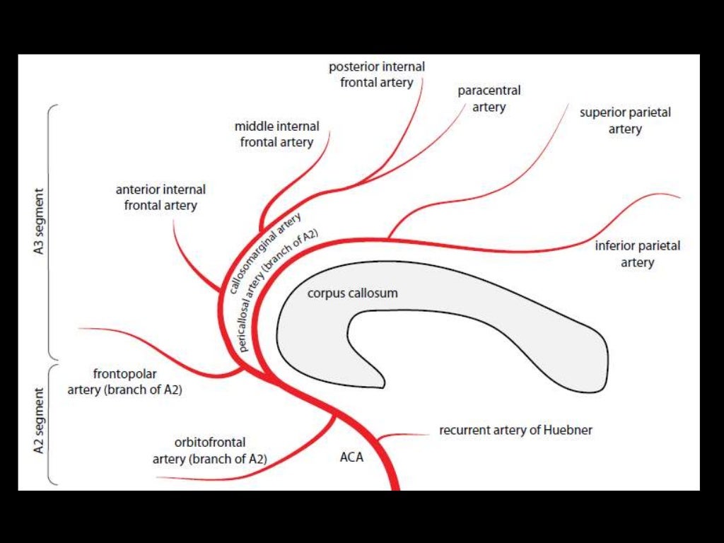
A1 is the segment originating from the internal carotid artery and extending to the anterior communicating artery. A2 extends from the anterior communicating artery to a region between the rostrum and genu. The A3 segment courses around the genu to the rostral part of the body. A4 and A5 segments are the continuation of the pericallosal artery. 

A1: ICA bifurcation to Acomm (horizontal)
A2: Acomm to origin of CMA around rostrum/genu junction (vertical)
A3: Genu to body of CC (proximal pericallosal)
A4/A5: Continuation of distal pericallosal (directly posterior)

(1) MCA (M4) , (2) MCA (M3) , (3) MCA (M2) , (4) MCA (M1) , (5) LT ICA , (6) LT
MCA , (7) Basilar artery , (8) PCA

NB angiographic sylvian point = apex of fissure sf. skull sylvian point a surface marking for fissure 3cm behind superior portion of zygomatic process


anteriorcerebralartery.jpg

Branches

A1
Medial lenticulostriate A.
AComm A.

A2
Recurrent artery of Hubner (= medial striate A.). Supplies head of caudate and anterior limb of IC. (Can be A1 branch)
Orbitofrontal A.
Frontopolar A.

A3
Callosomarginal A. (in cingulate sulcus)
Pericallosal A.
Anterior (internal) frontal A.
Middle (internal) frontal A.
Posterior (internal) frontal A.
Paracentral A.


NB Striatum =  Caudate + IC + lentiform nucleus (which = putamen + GP).

a) Deep (Perforating) Branches :
-Arise from the superior surface of the M1
segment
-They are grouped as the medial & late...

-2D frontal view following left ICA injections ,
these images show an aneurysm in the region
of the left MCA bifurcation/t...


*Arteries to the Parietal & Occipital lobes:
-These run posterior to the sylvian fissure , from
superior to inferior :
1-A...

1-Orbitofrontal , 2-Prefrontal , 3-Precentral , 4-Central , 5-Anterior parietal , 6-Post
parietal , 7-Angular , 8-Occipito...

Sunday, 2 December 2018

Monday, 5 November 2018

Identifying the Motor Strip


1. Surface anatomy

Taylor-Haughton Lines

Image result for taylor haughton lines
EAM just behind condylar process of mandible
Sylvian Fissure line starts at orbitotemporal angle- where the eyebrow ends.

  • The superior aspect of the motor cortex is almost straight up from the EAM near midline.  
  • 4-5cm posterior to coronal suture in adults. 
  • Or 2cm behind midpoint of nasion to inion. 
2. Radiological anatomy 

Surface anatomy of  the brain
The hand knob representing the hand motor area is in the pre-central gyrus. Looks like upside down omega. Delineates motor strip. 

Surface anatomy of  the brain


The motor strip typically terminates in the paracentral lobule, the pars bracket sign (arrowed) demarcates the posterior margin of the paracentral lobule.

Surface anatomy of  the brain

The thickness sign is simply that pre-central sulcus is thicker than post central- usually by 1.5:1. 

The postcentral sulcus is also usually bifid. 

The L sign- the superior frontal sulcus and precentral sulcus form an L shape. (See image at bottom) 

The IFG demonstrates an M shape - this is useful for Broca's area. It also follows that the pre-central gyrus is behind this. 

The hook sign represents the hand knob in sagittal plane and so indicates the motor strip. 

The pars marginalis sign follow the cingulate sulcus in midline sagittal view. The marginal sulcus (PM) is an extension of this. The post-central gyrus is in front of it.  

Image result for central sulcus sagittal

This is easy to use. Central sulcus is in red. Use it! 


Sunday, 7 October 2018

Trigeminal Ganglion Balloon Rhizotomy Procedure 

Background: 

WITHIN CRANIUMï‚—Middle meningeal nerve- travels with middle meningeal artery- supplies duramater


FO: foramen Ovale. TI Trigeminal impression (Meckel's gave is a dural recess which resides within this along the petrous apex). 

Position: Supine, neck hyper-extended (20 degrees) and rotated 20 degrees to contralateral side. Gel roll seldom necessary.  

Modified submental XR (draw trajectory vectors 2.5cm in front of tragus at inferior border of zygoma and mid pupilary line. Connect vectors to incision mark. Vectors also guide XR direction though there is a radiographers protocol) 

Entry Point: c. 3cm lateral to mouth

trigeminal nerve block

Ensure no penetration into mouth (double gloves)

Trajectory/Target: Aim for middle of FO

trigeminal nerve block

Will feel give on puncturing foramen ovale and should see CSF in primary cases. Caution- too lateral may enter temporal lobe and too medial lacerum (carotid). Don't plunge too deep either- just penetrate. 

trigeminal nerve block

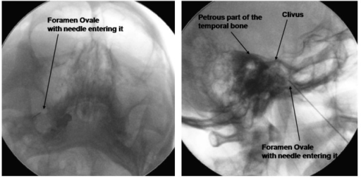
Can also identify petrous bone on submentatal XR (key XR landmarks are mandible, maxillary sinus, zygoma and petrous ridge). 

Once in, switch to lateral view. 

Submental View (With a 5 °Oblique Tilt) of the Foramen Ovale and Lateral View to Confirm the Depth of Needle Insertion


Inflate balloon slowly with approximately 0.8mls of dye. Radiograph should look pear shaped (by 0.5-0.7mls) and bradycardia expected. Don't inflate more than 0.8mls or ballon will burst. 
Dumbbell also acceptable, but sphere or irregular not (deflate and reposition). 
NB distance from needle tip to mid balloon 14-17mm (use a steristrip as a safety guard). 


A is incorrect (too high- up against sellar floor) B is correct and pear shaped. 

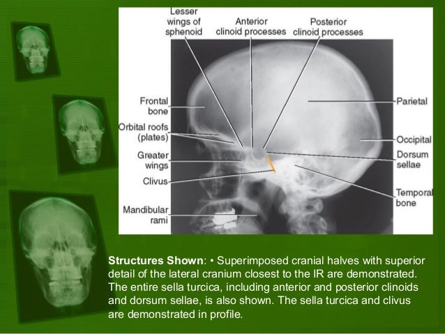
 Image result for lateral skull radiograph anatomy


1 and 6 pear shapes, 4 dumbbell. Others unacceptable. 

Complication avoidance: 
Diplopia most likely if balloon is too cranial (up towards sellar floor) or outside Meckel's cave (not pear shaped). 

Regional anatomy: coronal section
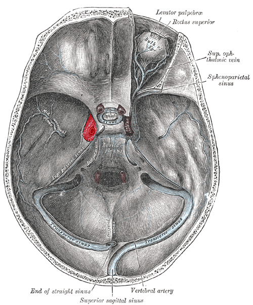
Image result for trigeminal balloon pear


Wednesday, 28 February 2018

CSF Cisterns

Quadrageminal is Supratentorial
Ambient is Supra and infra-tentorial
Inter-peduncular (between cerebral peduncles of midbrain) and Pre-pontine are infratentorial